Monday, November 7, 2011

Tinea pids ( Athlete's foot ) -Symptoms, treatment and prevention

Whate is tinea pedis (athlet's foot ) ?
         
           Tinea pedis is a fungal infection of the foot, that is common among homeless populations.Fungi are plant-like organisms tha live as parasites or saprophytes
           Fungi that cause superfcial infections of the skin and nails are called dermatophytes.
These fungi infect the keratin of the top layer of the epidermis as well as the nails and are responsible for Tinea pedis.
           Dermatophytes grow will in moist, occlusive environment. Condition such as diabetes and HIV / AIDS interfere with the body's immune functions and increase the risk of aquiring dermatophyte infection.

Whate is the prevalence of Tinea pedis ?
          
           Nearly everyone in the wold is exposed to the fungi tha cause Tinea pedis. Each person's immune system determines wherther infection results from such exposure. As adults age, tiny vravks develop in the skin of the feet, increasing the susceptibility to Tinea infections.
Once acquired , a fungal infection can linger inactively for years and later become active when person reaches the age of 60 - 70.
More than 70% of the US population will have Tinea pedis at the same time in their lives.
Diabetes is a significant risk factor, as diabetics are 50% more likely to have a fungal infection than non-diabetics.
         
How can i know that i've Tinea pedis ?
        There are three forms of Tinea pedis:
  • Interdigital - macerated, scaly, fissured skin occures between the toes, especially in the web space between the 4th and 5th toes.
  • Planter ( moccasin foot ) -fine, powdery scale is present on reddened background of the sole, heel, and side of the foot.
  • Vesicular (bullous) - an acute inflammatory reaction consisting of vesicles and pustules
Individuals may be asymptomatic or may experience burning, itching, or stinging.
        The diagnosis of Tinea pedis is usually made clinically and based upon the examination of the affected area.  Definitive diagnosis is made by scraping the skin for a KOH preparation, a skin biopsy, or culture of affected area.  The KOH is less likely to be positive in sever cases with maceation of the skin. 
 In mild cases the fungi can usually be recovered in a scraping.  In sever cases it is recovered less than half the time.
        In evaluation and diagnosis of Tinea pedis, clinicians should keep in mind that superinfection with bacteria can occure.
        Toenails infected appear thicker-ened, discolored, and dystrophic.

How can we treat Tinea pedis infections ?                    
        Treatment modalities come in several forms.
Topical agents include creams, powders, and sprays.  Creams and sprays are more effective than powders.
        Topical agents are generally effective in mild forms of interdigital Tinea pedis.  Non-prescition, over-the-counter (OTC) topival drugs work moderately well.  The less expensive OTC creams, such as clotrimazole (Lotrimin) and miconazole (Micatin), work as well as the more expensive OTC creams, but they require 4 weeks of traetment compared to 1-2 weeks needed with terbinafine (Lamisil) creams.  
In our practices, the first choice of treatment is usually an OTC cream.
        If Tinea pedis is extensive, a prescription topical antifungal would be used.  Econazole (spectazoe) and nystatin (Mycostatin) are prescription topical antifungal which are fungicidal and achieve a cure with shorter time.   In more extensive Tinea pedis or with failure of topical agents, oral medication by prescription can be used as fluconazole (Diflucan capsule)  
        The length of tratment with topical agent may take 4 weeks depending on the cream chosen.  Duration of therapy with oral agents can be 1-2 weeks depending on chosen medication.  
        In case of Tinea pedis with inflammatory signs and symptoms (including erythema, pruritis, and burning), a combination steroid/antifungal cream can be used.
         Onychomycosis requires treatment with oral antifungal medication for an extended period of time.

Mode of transmission of Tinea pedis

          Warm moist areas are fungi friendly.  Optimal environments are dark, damp, and warm.  Poor hygiene, closed footwear, minor skin or nail injuries, and prolonged moist skin creat ideal environments for transmission.
          Tinea pedis is contagious and spread through direct contact with people or opjects such as showers, shoes, socks, lockers rooms, or pool surface.  Pets can carry the fungus and may also be a source of transmission.

How can we prevent Tinea pedis infections
          Keeping the feet clean and dry is one of the best methods of prevention.  Other methods are well- ventilated shoes that fit properly and are not tight.
Alternating shoes daily will allow shoes to dry throughly in between wearing.  Wool socks draw moisture away from feet and are highly recomended.
Wearing sandals or flip-flops in public showers or pool area may also be helpful.
The use of foot powders is helpful for people susceptible to Tinea pedis who have frequent exposures to areas where the fungus is suspected.


Saturday, November 5, 2011

Prevention of atheroma – atheroma diagnosis, classification, prevention and drugs used in treatment

Atheroma definition and description
 Ath-er-o-ma is adeposit or degenerative accumulation of lipid-containing plaques on the innermost layer of the wall of an artery.
Atheroma is the most common cause of ischemic heart diseases, stroke and peripheral vascular diseases.
Family history of myocardial infarction confers an increased risk of ischemic heart diseases. Geneic factor is also important in the development of atheroma.

Pathophysiology
Atheromatous plaques are focal lesios of large and medium sized arteries. Atheroma start as fatty streaks in the intima and prgress to proliferative fibro-fatty growth that can protrude into the vascular lumen and limit blood flow.
These plaques are rich in both extracellular and intracellular cholesterol. During they development, they do not initially give rise to symptoms, but they may cause angina pectoris, intermittent claudication (clinical diagnosis gives for muscle pain ) or other symptoms depending on their anatomical location. They may rupture or ulcerate, in which the subintima acts as focus for thromboss , platelet-fibrin thrombi propagate and can occlude the artery, causin myocardial infarction or stroke.
Epidermiological observation have shown that  there is a strong positive relationship between the concentration of circulation cholesterol, specifically of low-density lipoprotien (LDL) fraction, and the risk of atheroma.
This relationship is non-linear and depends stongly on the presence or absence of other risk factors, including male sex, arterial hypertension, cigarette smoking, diabtes mellitus and left ventricular hypertrophy.
Cholesterol is mobilized from tissues in form of HDL particles. These are not atherogenic - indeed, epidemiological studies have identified HDL as being stromgly negatively correlated with coronary heart diseases.
There is a closed relationship between apolipoprotein A (Apo-a) and plasminogen linking atherogenesis to thrombosis.
Apo-a is present in a lipoprotein A (LP-A). The plasma concentration of LP-A varies over
a 100-fold range and is strongly genetically determind.
Most drugs have little efect ( nicotinic acid is an exception)
APO-A contains multiple repeats of one of the kringles of plasminogen ( kringle is a doughnut-shaped loop of amino acid held together by three internal disulphide bonds )
This leads to interference by LP-A with the function of plasminogen, which is the precursor of the endogenous fibrinolytic plasmin, and hence to a predisposition to thrombosis on atheromatous plaques.
Atheroma is classified to 8 types
  • Type I    : Isolated macrophage from cell
  • Type II   : Multiple from cell layers.
  • Type III  : Preatheroma, intermediate lesion.
  • Type IV  : Atheroma.
  • Type V   : Fibroatheroma.
  • Type VI  : Fissured, ulcerated, hemorrhagic, thrombotic lesion.
  • Type VII :Calcific lesion
  • Type VIII: Fibrotic lesion
Prevention and tratment of atheroma
Modifiable risk factors are potentially susceptible to therapeutic intervention.
These include smoking, obesity, sedentary habits, dyslipidaemia, glucose intolerance and hypertension.
Disappointingly hope, that hormone replacement treatment of post-menopausal woman would prevent atheromatous diseases were disproved by randomized controlled trials
* SOMKING *
Cigarette smoking is strong risk factor for vascular diseases. It causes vasoconstriction via activation of the sympathetic nervous system and platelet activation / aggregation with a consequent increase in thromboxan A2 biosynthesis, although the mechanism whereby smoking promotes atheroma is unknown.

* DIET AND EXERCISE *
Obesity is incresingly common and is an important risk factor, partly via its association with hypertension. Treatment is notoriously difficult.
Sedentry habit is a risk factor and regulare exercise reduce cardiovascular risk by reducing systemic blood pressure and increasing HDL.

* DYSLIPIDEMIA *
Most patient with dyslipidemia have a combination of genetic and dietary factors. Reducing the total plasma cholesterol concentration reduces the risk of coronary heart diseases and can cause regression of atheroma.

Dietary advice focuses on reducing saturated fat and correcting obsity rather than reducing cholesterol intake.

In people without clinical evidence of atheromatous disease, the decision as to whether to initiate drug tratment at any given level of serum lipids should be informed by the risk of coronary events.

* THREE MAIN CLASSES OF DRUGS TO TRAT  DYSLIPIDAEMIA *

*Statins*
Simvastatin, pravastatin, atorvastatin and rosuvastatin are available all over the world.
Randomized controlled trials have shown tha simvastatin, atorvastatin and pravastatin reduce cardiac events and prolong life, and safe.

Pravastatin is distributed selectively to the liver and is tolerated even by some individuals who develop myalgia on other statin, but is less potent.

Rosuvastatin lacks clinical end-point data,  but is more potent.

Cerivastatin is highly potent statin. It was withdrawn because of rhabdomyolysis ( a rapid destruction of skeletal muscle resulting in leakage into the urine of muscle protien myoglobin)
and drug interaction.

*DRUGS USED TO REDUCE CHOLESTEROL ABSORBTION *
   * EZETIMBE *
It is most often used in compination with diet and statins for sever hypercholesterolaemia; also in occasional patient who cannot tolerate statins or where statins are contraindicated, and in rare cases of homozygous sitosterolaemia (a rare autosomal recessively inherited lipid metabolism disorder

Diarrhoea, abdominal pain or headache are problems. It is contraindicated in breast-feeding.

* ANION-EXCHANGE RESINS *
Colestyramine or colestipol were used for hypercholesterolaemia, but have almost completely superseded by statins.
Resins retained an important nich as addd-in treatment in sever disease which was inadequately responsive to statin monotherapy.
This role has now been taken by ezetimbe which is effective and well tolerated.

They cause malabsorption of fat soluble vitamins and interfere with the absorption of many drugs.

* FIBRATES *
Benzafibrate, gemfibrate and febofibrate are used mainly for patient with mixed dyslipidaemia with severely raised triglycerides especially if they are poorly responsive to statins.

Colfibrate, wich was used in a World Health Organization (WHO) trial, is less often used because it increases biliary cholesterol secretion and predisposes to gall-stone.

Furthermore, while it reduced the number myocardial infarction in the WHO trial, this was offset by an increased number of cancers of various kinds.

The studies have shown that fibrates have a marked effect in lowering plasma triglycerides with a modest reduction in LDL and increase in HDL.

Fenofibrate has an additional uricosuric effect.


Friday, November 4, 2011

DiGeorge syndrome - causes , symptoms and complications


Digeorge syndrome ( dgs)
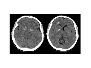
Brain computer tomography cuts of the patient,
demonstrating basal ganglia and periventricular calcification

What is Digeorge syndrome ???
DiGeorge syndrome is a disorder caused by a defect in chromosome 22, which hinder the development of several body systems.
Before that tremendous progress in genetics, Digeorge syndrome was known by many names like
Shprintzen syndrome, conotruncal anomaly face syndrome, Strong syndrome, congenital thymic aplasia, and thymic hypoplasia .
Now , "22q11.2 deletion syndrome" is frequently used which is more accurate , but old names are still used
How does DiGeorge syndrome occur   ?
Everyone has two copies of any chromosome . any chromosome contains 500-800 genes . DiGeorge syndrome happens when a part of chromosome 22 is deleted.
In fetus who has DiGeorge syndrome one copy of chromosome 22 is imperfect and so about 30-40 genes are missed .That part is known as 22q11.2.
That process occurs randomly whether in the mother egg or in the father sperm, or it may be early during baby development.
symptoms and signs of DiGeorge syndrom
Some signs and symptoms may be apparent at birth, but others may not appear until later in infancy or early childhood.Signs and symptoms of DiGeorge syndrome can vary significantly in type and severity.This variation depends on what body systems are affected and how severe the defects are.
This may include: Failure to thrive, Failure to gain weight, Poor muscle tone, Shortness of breath,Twitching or spasms around the mouth, hands, arms or, Frequent infections, Difficulty feeding , Delayed development, such as delays in rolling over, sitting up or other infant milestones, Delayed speech development, Learning delays or difficulties, A gap in the roof of the mouth (cleft palate) or other problems with the palate, Certain facial features, such as low-set ears, wide-set eyes or a narrow groove in the upper lip
But how can DiGeorge syndrome be diagnosed ?
Diagnoses of DiGeorge syndrome is based mainly on laboratory tests.
Some heart deects are common in DiGeorge syndrome, so presence of these defect may prompt the doctor to order a laboratory test to chromosome 22.
DiGeorge syndrome and complications
So many complications can occur according to the role was supposed of that deleted part in baby body system. We cannot expect these complications, but common one are:
* Cleft palate*
Cleft palate is an open in the roof of the mouth, so can make the patient suffers from difficulty in swallowing.
* Thymus gland dysfunction *
In children with DiGeprge syndrome thymus gland may be small or missing wiche resulting in poor immunity and frequent sever inections . HOW?
The thymus gland in children, located beneath the breastbone, is where T cells — a type of white blood cell — mature. Mature T cells are needed to help fight infections. As you grow older, the thymus decreases in size, and other parts of your body take over the role of the thymus in the immune system.

* Hypoparathydoidism *
Parathyriod hormone (PTH) is secreted by parathyroid gland located in our nick. That hormone is responsible for maintaining appropriate levels of calcium and phosphorous in our body. DiGeorge syndrome can result in smaaler parathyroid gland, so too little PTH is secreted.
Hypoparathyroidism results in low calcium level and high phosphorous level in the body.

* Heart deects *
A common comlication o DiGeorge syndrome that result in an insufficient supply of oxygen to the body systems.
Defects may include a hole between the lower chambers of the heart (ventricular septal defect); only one large vessel, rather than two vessels, leading out of the heart (truncus arteriosus); and a combination of four abnormal heart structures (tetralogy of Fallot)
* Autoimmune diseaes*
Having small or missing thyms gland, resuluts in high risk of autoimmune diseases like rheumatoid arthritis and Graves disease (http://en.wikipedia.org/wiki/Graves'_disease )
* Social promblems *
The part deleted of chromosome 22 may cause developmental and functional problem in brain.
Many children with DiGeorge syndrome suffering from autism and attention-deficit/hyperactivity disorder (ADHD). Delays in toddler speech development and learning difficulties are common.
When patient be older, he may suffer of mental disorders like depression, anxiety disorders, schizophrenia and other psychiatric disorders.

Is there any treatment for DiGeorge syndrome ?
Honestly, there is no cure for DiGeorge syndrome, only we can manage the symptoms such as :
Cleft palate & heart defects
they have to be surgically treated.
Thymus dysfunction
Treatment may require a transplant of thymus tissue, specialized cells from bone marrow or specialized disease-fighting blood cells if thymus gland is missed.
But in case of mild thymus dysfunction, frequent infections are treated as they would be in any child and they have to follow the normal schedule of vaccines. For most children with moderate thymus impairment, immune system function will improve as they grow older.
Hypoparathyroidism
Can be managed with calcium supplements, vitamin D supplements and a low phosphorus diet. If enough of the parathyroid tissue is intact, it's possible the child's parathyroid glands will eventually regulate calcium and phosphorus levels without a specialized diet.
At the end we have to know that if we have such a child, we should praise God and thank him for his test to admit us to his Paradise.

support them